How will ITRI's Point-of-Care AI-DR system address critical healthcare needs related to diabetic retinopathy and diabetic macular edema?
Currently, most medical imaging AI-assisted diagnosis systems are used to assist doctors in their respective specialties, such as AI assisting radiologists in X-rays and CT scans, AI aiding pathologists in pathology images, and AI supporting internal medicine doctors in ultrasound images. The purpose of the colour fundus image AI-assisted diagnosis and detection system is not to assist ophthalmologists in diagnosing eye diseases but to provide diabetes care physicians (e.g., those in metabolism and family medicine) with the capability to diagnose retinal diseases. Diabetic patients can have fundus examinations during their regular check-ups without the need to be referred to ophthalmology, saving them time and registration fees. This one-stop service speeds up the process of fundus examinations, leading to the early detection, early treatment, and the prevention of retinal diseases, which can cause significant burdens on families.
From a business perspective, what opportunities and challenges do you see in the adoption and commercialisation of AI-assisted diagnostic systems like Point-of-Care AI-DR in the global healthcare market?
Computer-Aided Diagnosis (CADx) provided by artificial intelligence needs to gain trust from doctors. If doctors cannot trust the results generated by CADx, they will not use such software. How do CADs earn doctors’ trust? Generally, trust arises when doctors understand the diagnostic logic and rules behind the equipment. Doctors evaluate the credibility of CADs-generated results by combining computer's logic and rules with their professional knowledge and experience. However, AI models constructed through extensive calculations do not provide logic and rules on their own. Therefore, through discussions with doctors, this project has confirmed that the severity classification of DR/DME is related to lesion and anatomical landmarks. For example, when fundus images only show microaneurysms and no other lesions, the severity classification of DR is Mild NPDR. When lesions such as HE (Hard Exudates) appear in the macular area, it indicates DME. As a result, this Computer-Aided Detection (CADe) provides four types of lesion classifications and two anatomical marker locations to increase the confidence of doctors to explain the severity of DR/DME to patients based on information such as lesion type, quantity, and location, thereby improving patient compliance with medical recommendations.
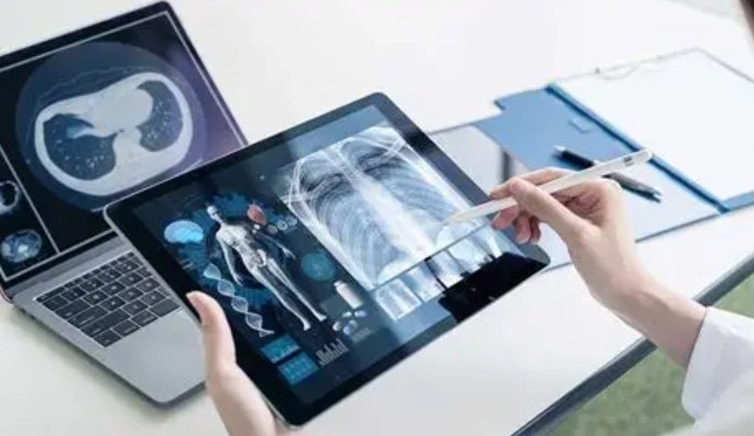
Currently, the world is facing the challenge of an ageing population and rapidly growing healthcare demands. However, healthcare systems have insufficient capacity to meet these increasing demands. Globally, there is a strong hope that AI can help accelerate the development of smart healthcare, increase the early detection rate of diseases, reduce misdiagnosis and missed diagnoses, and effectively manage the rapid growth of patient populations. This provides a significant opportunity for AI in the healthcare sector to address these pressing issues and enhance the overall quality and efficiency of healthcare services.
Can you share any insights into the collaborative efforts that went into the development of Point-of-Care AI-DR, both within ITRI and through potential partnerships with healthcare providers or industry stakeholders?
Medical data, according to personal data protection regulations, falls under the category of special personal private information. It requires approval from the hospitals’ Institutional Review Board (IRB) after a thorough professional review process. Our project began its engagement with the first medical centre in the fourth quarter of 2017. After discussing the details, quantities, and schedules of data collection with collaborating doctors, we drafted a research proposal. This proposal went through multiple rounds of review and adjustments based on feedback from the IRB, ultimately receiving IRB approval. In 2018, we obtained tens of thousands of fundus images from three medical centres.
Data preparation took two and a half years. Our team worked diligently, visiting over a hundred domestic ophthalmologists. In the end, we obtained contributions from more than 50 domestic ophthalmologists who provided their professional knowledge and expertise in manual annotation. This effort allowed us to accumulate a dataset that includes over 10,000 images with annotations, covering more than 20 types of eye diseases, lesions and anatomical markers. This dataset is currently the most comprehensive and extensive fundus image dataset globally. Our project involved calculating confusion matrices to evaluate the consistency of data annotations, whether among different physicians or within individual physicians. When consistency issues arise, we engage in discussions with multiple physicians or individual physicians to address concerns and clarify annotation issues. This process helped ensure that the annotation standards for diseases/lesions were appropriately adjusted and revised as needed.
The development of this technology requires collaboration with healthcare institutions and personnel. Therefore, gaining trust from hospitals, persuading them to release data, and effectively communicating with doctors and devising mutually beneficial strategies with physicians are important milestones in the development of this technology.
What are the future plans for ITRI's Point-of-Care AI-DR system, and how do you envision its impact on healthcare and the technology industry in the coming years?
In the field of AI-assisted diabetic retinopathy detection technology, the plan is to continuously expand its application scope and drive the service ecosystem. This involves forming strategic alliances and collaborations with fundus camera manufacturers, medical cloud service providers, PACS (Picture Archiving and Communication System) medical image management system providers, and other emerging service providers with new business models. The goal is to assist healthcare providers worldwide in improving the management of diabetic eye complications.
Considering that microvascular and macrovascular complications can lead to disability and death in over 500 million diabetic patients globally, the development of cardiovascular disease prediction plays a crucial role. The aim is to effectively predict and accurately diagnose cardiovascular diseases before they occur, allowing for appropriate intervention measurements. This comprehensive approach to diabetes care can reduce or delay the risks of these diseases and the associated medical expenses.
In summary, these efforts aim at improving healthcare outcomes, preventing complications, and ensuring better overall care for diabetic patients worldwide.
Ayesha Siddiqui