Scientists have developed tiny Three-dimensional (3D) -printed scaffolds that can guide the regrowth of nerve cells, possibly restoring the sense of touch and movement control in patients with the damaged peripheral nervous system.
Now a days 3D printing has practiced as the technology with more accurate and handy in all sectors.
3D scaffolds serve as temporary substrates for supporting and guiding tissue formation in various in vitro and in vivo tissue regeneration settings.
Researchers from the University of Saskatchewan in Canada are looking at how we can use 3D printing to help damaged nervous systems to regrow.
Our body beyond the brain and the spinal cord is controlled by peripheral nervous system. This may get damaged by poor diet, toxins, and trauma and diabetes. This damaged peripheral nervous system can ultimately affect our sense of touch and our motor control.
The current standard for treating large gaps in the nervous system due to damage is nerve autografts, where donor nerves from another part of the body are used to repair the damaged parts.
However, this process is not perfect; there are limited donor sites for nerve repair, and even successful grafts often only restore a portion of the nerve's original functionality.
In order to improve nerve regeneration, a combination of 3D printing and biotechnology may be the answer.
Researchers used Schwann cells, supporting cells in the nervous system that can force nerve cells to grow properly, in a 3D printed hydrogel-based scaffold in order to promote and guide the regeneration of the damaged nerves.
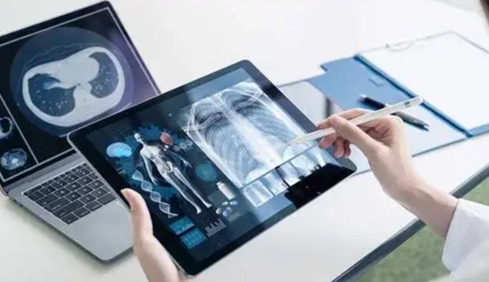
Conventionally, testing techniques like micro-CT imaging would be used to image samples like these bio-printed scaffolds. However, one of the steps in this testing method is to freeze dry the samples.
This freeze-drying process may partially damage the scaffolds, meaning that the structure that is seen from the images is not precisely the structure that the intact scaffolds would have. This imaging process can also take several hours to scan one sample.
The samples can be imaged without any freeze drying, preserving the true scaffold structure. The process is also much faster, taking only about 10 minutes to scan one sample.
Scientists want to work out a few problems in the scaffolds before this method sees regular medical usage. They want to try out some different techniques to better direct nervous system growth.