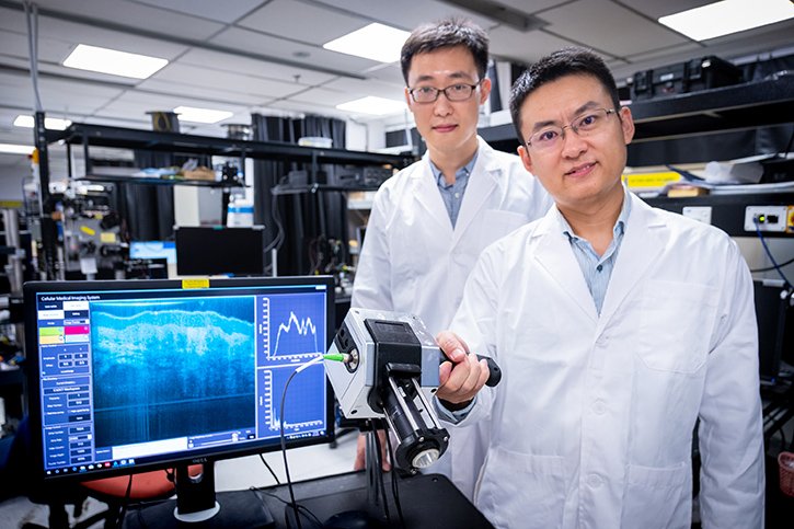
Singapore designs high resolution medical device
March 30, 2020 | Monday | News
The technology behind the device is a result of six years of optical imaging research

Image credit- NTU
Scientists at Nanyang Technological University, Singapore (NTU Singapore) have developed the prototype of a handheld medical imaging device that can produce images down to resolutions of 1 to 2 micrometres.
This is detailed enough to spot the first signs of tumours in specific cells and is about 100 times higher resolution than what X-Ray, computed tomography (CT) and Magnetic Resonance Imaging (MRI) machines can provide.
The technology behind the device is a result of six years of optical imaging research and was jointly developed by a team from NTU with researchers at the Harvard Medical School and the University of Alabama, U.S.A.
Relying on a new imaging technology known as micro ‘Optical Coherence Tomography’ (OCT), the device emits a spectrum of light between 700 to 950 nanometres, known as near-infrared light. This harmlessly penetrates human tissue and organs, and the device then measures the delay time of the ‘echo’ from its light waves as they strike different tissue structures. This information will then be used to construct cross-section images of what is being scanned.
The results are sent in real-time to a computer system running software developed at NTU, which assists in diagnosis by assembling the 2-D cross-section images into a three-dimensional picture and rendering different parts in colour.
Prof Liu and his team are conducting more in-depth research into OCT technologies, to further improve the device and extend industry collaboration with other healthcare companies.


