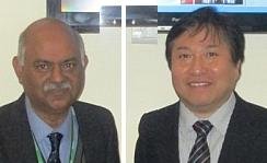
Dr R KGupta, director and HOD, radiology, Fortis, India (L), and Dr Taro Takahara, pioneer of diffusion weighted imaging with background suppression (DWIBS) from Japan (R) at the Fortis symposium
New Delhi: Senior radiologists from around the world deliberated the latest advances in magnetic resonance (MR) techniques in neurology and whole body imaging, at a recently held advanced international symposium, organized by the Fortis Memorial Hospital, India.
The event saw participation from eminent doctors, including Dr RK Gupta, director and head of the department, radiology, Fortis Memorial; Dr Taro Takahara, pioneer of diffusion weighted imaging with background suppression (DWIBS) from Japan; and Dr Ponnada Narayana, professor, University of Texas Health Science Center, US. Several others also showcased a unique combination of two cutting-edge whole body MR imaging technologies for the first time in the world, for the early detection of tumors.
This path breaking effort deploys m-Dixon and DWIBS, the two advancements in magnetic resonance (MR) that provide precision high resolution images of one millimeter thickness. This enables the sighting and diagnosis of diseased tissues, in their infancy, making it possible for targeted therapy solutions to be applied early.
Highlighting the advantages of this new innovation, Dr Gupta, said that, "Cancer treatment can be more effective if the disease is diagnosed and treated in its infancy much before the physical symptoms manifest themselves and the disease reaches an advanced uncontrolled state. The fusion of the two cutting edge technologies m-DIXON and DWIBS, in advanced MR application enables the detection of tiny tumors. One millimeter slicing has been the gold standard in MR investigations, as it provides high resolution images. With its application on the whole body, tumors or infections that were not revealed in MR imaging earlier, can now be detected easily in as little as 20 minutes and without exposing the patient to radiation."
Dr Gupta further said, "Many times patients who have successfully battled cancer may suffer from remission and other systemic infections like tuberculosis making early diagnosis difficult. With the help of whole body MR imaging we are now able to detect and pinpoint the exact cause of the ailment. Naturally, this accelerates the treatment protocols."